Digital X-Rays for Dogs & Cats
X-rays use electromagnetic waves to create an image of the body’s internal structures. They’re especially useful in identifying diseases in the chest (asthma, bronchopneumonia, cancer, and fluid build-up from congestive heart failure, for instance) along with musculoskeletal diseases including arthritis, developmental problems such as hip dysplasia, fractures, and cancerous bone lesions. They can also be useful in evaluating the abdomen (identifying urinary bladder stones, a tumour, problems such as constipation and ‘megacolon’, and so on).
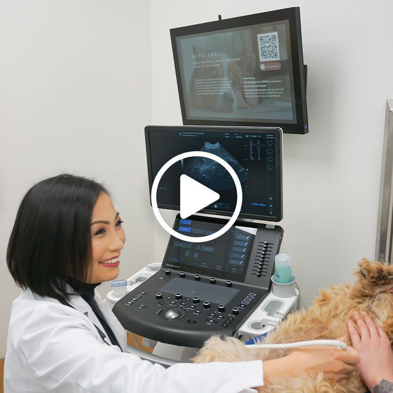
Cat & Dog Ultrasound
Ultrasound uses high-frequency sound (ultrasound) waves to produce images of structures within the body. It involves no radiation whatsoever. Next to X-rays, ultrasound is the most common imaging method used in veterinary medicine. It allows us to see the abdominal organs in more detail, including the bladder, kidneys, liver, spleen, stomach, and intestines. It also allows us to assess the structure of the heart for abnormalities in muscle walls and valves, which is important if we’re considering a procedure involving sedation or general anesthesia in a patient with a heart murmur, for example. With ultrasound, we can also look for fluid build-up in the chest or abdomen and safely guide fluid and tissue sampling for further analysis.
Sedation is used only if a patient is too wiggly or experiencing too much pain to tolerate the mild pressure of the ultrasound probe.
We Take Our X-rays "Hands Free"
Taking an X-ray traditionally requires two technicians to properly position and restrain a pet to capture an image. To avoid radiation exposure, our techs use hands-free techniques – positioning devices, quiet, calm handling, and if needed, some mild sedation to help a patient relax so they can capture an image of high diagnostic quality. Your veterinarian will inform you if a light sedative is needed and will choose one that’s tailored to your pet.
There are certain X-rays (for example, those evaluating painful orthopedic conditions) that require heavier sedation. Your veterinarian will let you know if that level of sedation is indicated for your pet.
For more information, visit: https://handsfreexrays.com/.
